Figure 4.

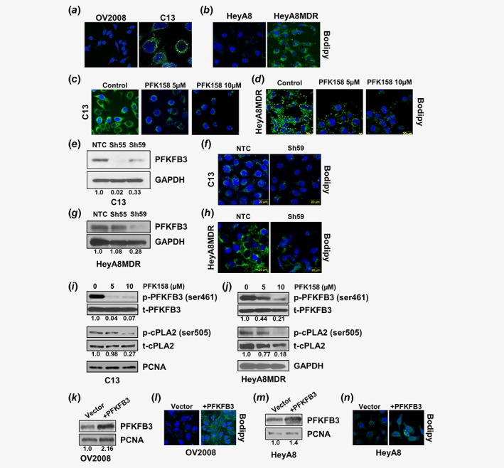
Inhibition of PFKFB3 triggers degradation of LDs. Chemosensitive (OV2008, HeyA8) and chemoresistant (C13, HeyA8MDR) cells were labeled with Bodipy 493/503 (green) and DAPI (Blue) to determine the cytoplasmic LDs (a‐b) and nuclei, respectively. Bodipy staining performed in C13 (c) and HeyA8MDR (d) cells to detect LDs in PFK158 (0‐10 μM) treated cells. Bodipy staining performed to assess LDs after transient downregulation of PFKFB3 (confirmed by western blot; e, g) in C13 (f) and HeyA8MDR (h) cells. Protein expression of p‐PFKFB3, t‐PFKFB3, p‐cPLA2, t‐cPLA2 and GAPDH are detected by western blotting in PFK158 treated (0‐10 μM) C13 (i) and HeyA8MDR (j) cells. Bodipy staining of LDs in OV2008 (l) and HeyA8 (n) cells after transient transfection with either empty vector or PFKFB3 (verified by western blot) (k, m). [Color figure can be viewed at wileyonlinelibrary.com]
