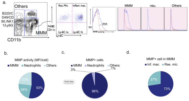
Fig. 1. Myeloid cells are the main source of matrix metalloproteinase (MMP) activity in EAE.
a Cell surface markers used to subdivide cells into various subsets of leukocytes in the brain of saline-treated control EAE mice 12 days after immunization. Numbers denote percent MMP-positive cells in the saline (34%± 5%) group (Red line: percent MMP-positive cells in sham mice; Blue line: percent MMP-positive cells in EAE mice). b Flow cytometric analysis revealed MMP activity mainly derived from microglia/monocytes/macrophages (MMM) in brains of EAE mice (53%). c MMP mean fluorescence intensity (MFI) per cell for different cell types in brain, as identified by flow cytometry. MMM cells contribute to most of the MMP activity (96%). d The percentage of MMP positive cells in inflammatory macrophages and resident microglia, 73% and 27% respectively. SSC = side scatter; neu. = neutrophils; MMM =microglia/monocytes/macrophages; MFI = mean fluorescence intensity; Inf. mac = inflammatory macrophages; Res. Mic = resident microglia

