
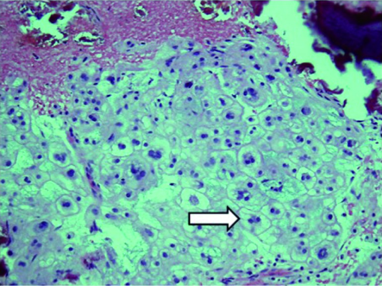
Figure 3.
Histology from T6 chordoma resection demonstrating (arrow) the physaliferous cells (soap-bubble appearance) found in chordomas. H&E staining using a ×20 objective lens.
Official websites use .gov
A
.gov website belongs to an official
government organization in the United States.
Secure .gov websites use HTTPS
A lock (
) or https:// means you've safely
connected to the .gov website. Share sensitive
information only on official, secure websites.
Histology from T6 chordoma resection demonstrating (arrow) the physaliferous cells (soap-bubble appearance) found in chordomas. H&E staining using a ×20 objective lens.