
Figure 4. Left-Lateralized Frontoinsula Atrophy Predicts Diminished Parasympathetic Activity and Prosocial Behavior Deficits.
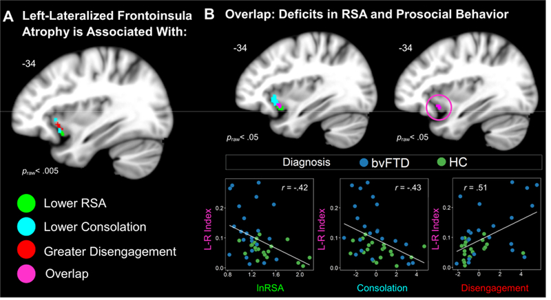
(A) Left-greater-than-right FI atrophy correlated with lower RSA, lower consolation behavior, and greater disengagement (praw< .005). (B) Lowering the threshold (praw< .05) revealed an overlapping cluster (violet) where voxel intensity significantly correlated with RSA, consolation, and disengagement. We extracted the gray matter volume from this cluster in each participant. Scatterplots are shown to illustrate the independent associations that left-lateralized frontoinsula atrophy had with all three measures. Statistical maps of the lateralization results are superimposed on the left hemisphere of the Montreal Neurological Institute template brain. Maps represent T-scores at praw< .005, uncorrected (T> 2.70) and praw< .05, uncorrected (T> 1.68). Frontoinsula (FI), left (L), respiratory sinus arrhythmia (RSA), right (R).