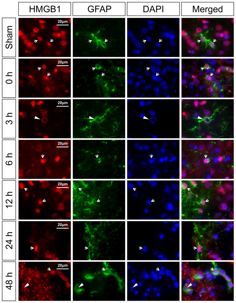
Fig. 4. Immunohistochemical double staining of HMGB1 with astrocyte marker (GFAP) in the neonatal rat brain cerebral cortex at zero, 3, 6, 12, 24, and 48 h after exposure to HI injury.
The HMGB1 expression was mainly in the nucleus of astrocytes in the sham-operated, and at zero, 3, 6, 12, and 24 h after HI. HMGB1 translocation and release was not observed in the astrocytes. DNA damage with ring-like structures co-localized with HMGB1 was observed in some astrocytes (3 h, white arrowhead). Furthermore, some astrocytes showed negative nuclear staining of HMGB1 at 48 h after HI injury (48 h, white arrowhead). White arrows show the representative nuclear localization of HMGB1 in GFAP positive cells. Scale bar = 20 μm.

