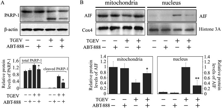
Fig. 2.
The effect of ABT-888, a PARP-1 inhibitor, on the translocation of AIF. (A) PK-15 cells were incubated with 20 µM ABT-888 for 1 hr and subsequently infected with TGEV for 36 hr. Cells were collected and then subjected to Western blot analysis for PARP-1. The total protein and cleaved protein levels of PARP-1 were calculated by densitometry of the corresponding bands after normalization to β-actin. (B) Cells were treated as in (A). The mitochondrial and nuclear proteins were extracted from the collected cells, and then subjected to Western blot analysis. AIF protein expression in the mitochondria and nucleus was calculated by densitometry of the corresponding bands after normalization to Cox4 and Histone H3, respectively. (C) All data are means ± SD of triplicates from three independent experiments. *P<0.05, **P<0.01 versus TGEV infection without ABT-888.

