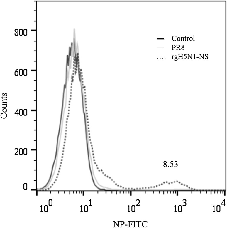
Fig. 3.
Flow cytometry for determining infectivity in THP-1 monocytic cells infected with NS reassortant PR8 virus. THP-1 cells infected with PR8 virus or rgH5N1-NS virus at m.o.i. of 3. At 24 h post infection, cells were analysed for influenza NP by flow cytometry. Numbers indicate percentages of cells in each gate. Uninfected cells are used as control staining. Histogram shows the average percentage (mean ± SD) of NP+ THP-1 cells from 3 independent experiments (two-tailed t test: P = 0.010)

