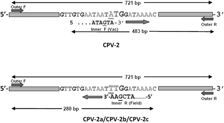
Fig. 1.
Schematic representation of CPV ARMS-PCR strategy for differentiating vaccine (CPV-2) and field strains (CPV-2a/CPV-2b/CPV-2c) showing primer binding region and amplified product formed. The bold case (bigger font size) represents the target codon (position 259–261) and primer binding site. Deliberate mismatch of the nucleotide at − 2 from 3′ end position within primers is underlined

