
Figure 2.
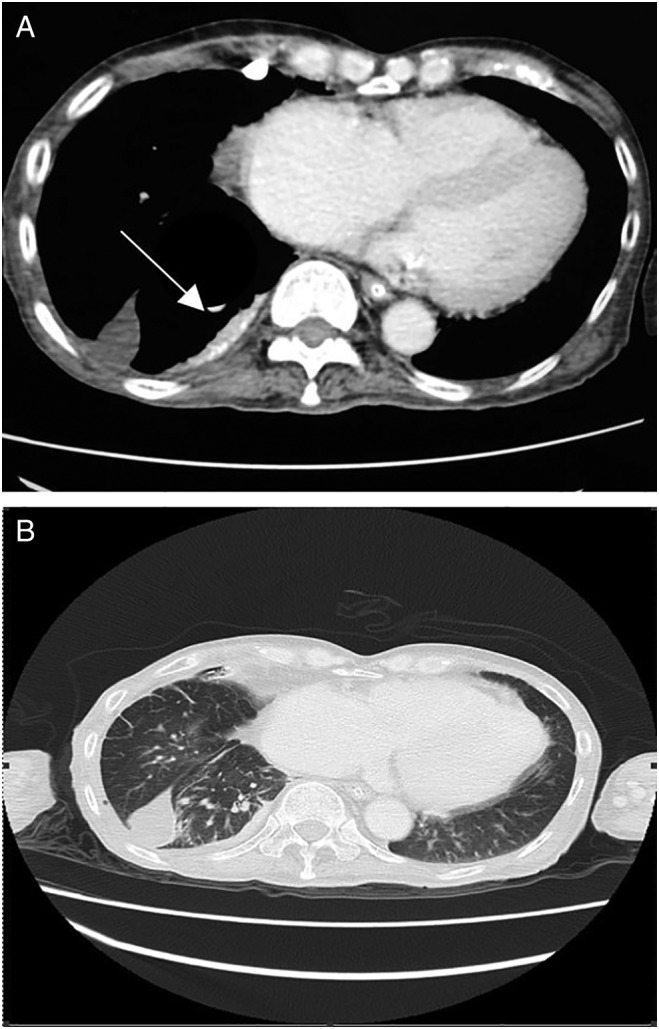
(A) Contrast‐enhanced computed tomography (CECT) thorax showed a 1.4 cm thick focal pleural plaque (white arrow) with calcification at the medio‐posterior aspect of right lower lobe. (B) Loculated fluid in the horizontal fissure and interlobular septal thickening.