Fig. 1.
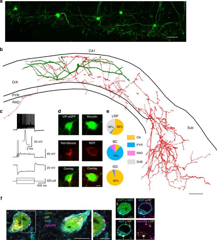
Identification of VIP-LRPs in the VIP-eGFP mouse. a Two-photon image (maximal projection of a z-stack of 200 µm height) of the CA1 area from an acute hippocampal slice (300 µm) of a VIP-eGFP mouse showing the location of GFP cell bodies, axons and dendrites in the O/A area of CA1. Scale bar: 100 µm. b Reconstruction (the axon is shown in red, the dendrites are shown in green) of a VIP-LRP cell that was recorded and filled with biocytin in a slice obtained from a VIP-eGFP mouse. Scale bar: 100 µm. c Representative voltage responses of a VIP-LRP to hyperpolarizing (−240 pA), and depolarizing (+80 pA and +280 pA) current injections, with an inset illustrating the first spike evoked by +80-pA current pulse. d Confocal images showing RetroBeads labelling of a VIP-LRP soma (left) after injection in subiculum and immunoreactivity for M2R in a VIP-positive neuron labelled with biocytin (single focal plane, right). Scale bar: 10 µm. e Pie charts illustrating the mean axonal distribution in different layers (based on axon length obtained following reconstruction in Neurolucida) for groups of cells corresponding to three different cell types: VIP-LRP (n = 10), VIP-BC (n = 5) and IS3 cell (n = 6). OA oriens-alveus, PYR stratum pyramidale, RAD stratum radiatum, SUB subiculum. No axon was detected within stratum lacunosum moleculare (LM) for the three cell types. Statistically significant differences in the axon distribution between VIP-LRP and VIP-BCs, VIP-LRP and IS3, and VIP-BCs and IS3 at **p < 0.01, one-way ANOVA followed by Tukey’s test. f VIP-LRP cells, identified by somato-dendritic membrane M2R immunoreactivity (blue), are innervated by terminals rich in presynaptic mGluR8 (purple). Single optical slices (0.45 mm thick) of confocal images of quadruple immunoreactions as indicated: i, right, framed area at higher magnification; ii, four VIP+ terminals (arrows) show mGluR8 immunoreactivity. Scale bars: 10 µm (left), 5 µm (middle), 5 µm (right)

