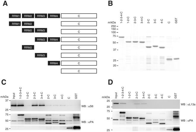
Figure 3.
Localization of ribosomes on PABP. (A) A schematic representation of PABP and RRM-deletion mutants. (B) C-terminally His-PA–tagged wild-type PABP (1-2-3-4-C), RRM-deletion mutants and GST proteins (1 μg each, Coomassie brilliant blue stained). (C) and (D) Binding of the 40S ribosomal subunit (C) and the 60S subunit (D) to PABP (1-2-3-4-C) or its deletion mutants immobilized on the TALON resin. Input: one twentieth of the ribosomal sample was analyzed at the same time.

