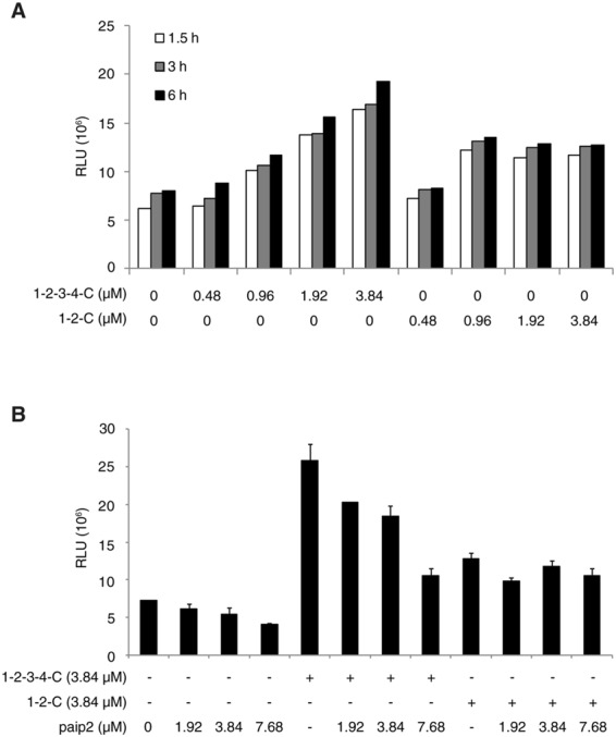
Figure 6.

Functional analysis of PABP interaction with the ribosome. (A) Cap-Rluc-A RNA (0.1 μM) was translated in the reconstitution system in the presence of increasing concentrations (0 to 3.84 μM) of PABP (1-2-3-4-C) or a truncated PABP (1-2-C). At indicated times, an aliquot of each sample was removed for the Rluc assay. Each bar represents the mean of two experiments. (B) Cap-Rluc-A RNA (0.1 μM) was translated in the reconstitution system with PABP or PABP (1-2-C) (3.84 μM each) in the presence of increasing concentrations (0 to 7.68 μM) of Paip2 for 6 h. After translation, Rluc activity was measured. Each column and bar represent the mean and standard deviation of three experiments, respectively.
