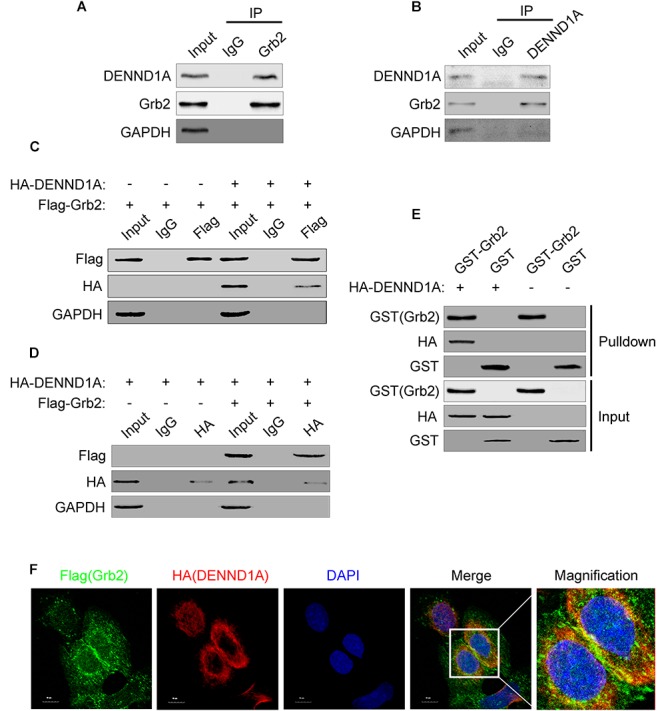
FIGURE 3.

DENND1A and Grb2 combine with each other. (A) BGC-823 cells lysates were immunoprecipitated with an anti-Grb2 antibody or (B) anti-DENND1A antibody, and then both unprocessed lysates (Input) and immunoprecipitates were analyzed by western blot with indicated antibodies. (C) HEK-293T cells transiently transfected with Flag-Grb2 and HA-DENND1A were immunoprecipitated with an anti-HA antibody or anti- Flag antibody, and then both unprocessed lysates (Input) and immunoprecipitates were analyzed by western blot with indicated antibodies. (D) GST-Grb2 was incubated in vitro with lysates of HEK-293T cells transfected with or without HA-DENND1A, and coprecipitation of DENND1A with GST-Grb2, bound to glutathione-beads, was analyzed by western blot. GST, GST alone used as a control. (E) BGC-823 cells expressing Flag-Grb2 and HA-DENND1A were cultured on coverslips and fixed and double-stained with anti-HA and anti-Flag antibody. (F) Original colors were Flag-Grb2, green; HA-DENND1A, red; and nuclei, blue. Merged pictures represent the composite of all channels, with yellow regions indicative of colocalization. Scale bar, 10 μm. All experiments were repeated at least three times.
