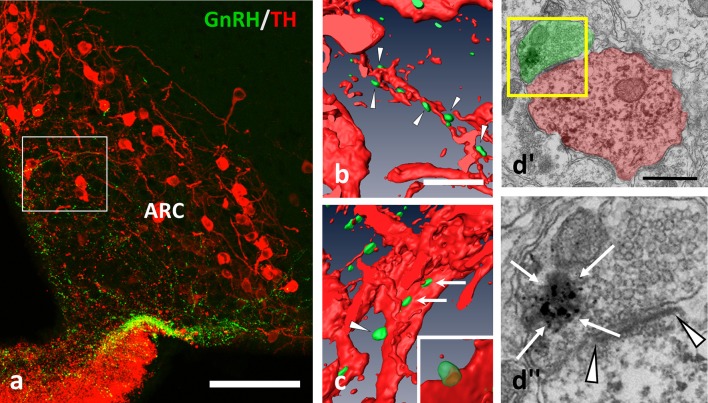
Figure 2.
GnRH-IR varicosities (green) establish multiple appositions on TH-IR neurons (red) also in the Arc. (a–c) Axo-dendritic and axo-somatic appositions are indicated by arrowheads and arrows; (b,c) are snapshots from the 3D reconstructed and rotated image of the boxed area in (a). The presence of asymmetric synapses (d; arrowheads) were confirmed also by the arcuate appositions. (d)” The GnRH-IR axon terminal contained both dense-core granules (arrows, heavily labeled with SGI-NiDAB) and round-shaped, small, clear vesicles, as shown in the high power image of the boxed area of (d)'. Scale bar in (a) is 100 μm; in (b) and (c) 10 μm; in (d') it is 500 nm.

