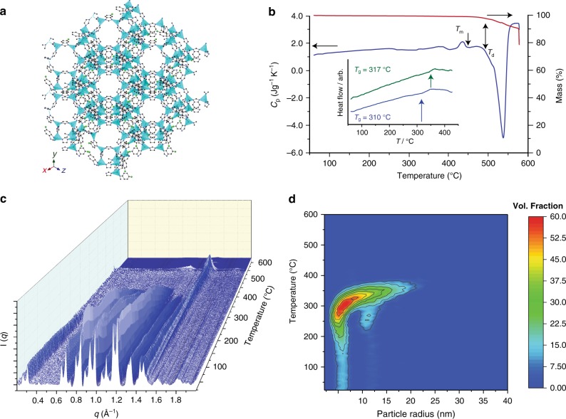
Fig. 1.
Liquid and glass formation of ZIF-76. a The structure of ZIF-76, as determined by single-crystal X-ray diffraction20. Zn light blue, Cl green, C grey, N dark blue, H omitted for clarity. b The isobaric heat capacity (Cp) and mass change (%) of ZIF-76 measured during a DSC-TGA upscan at 10 °C min–1, highlighting the stable liquid domain between Tm and Td. Inset shows glass transitions for agZIF-76 (blue) and agZIF-76-mbIm (green). c Temperature resolved WAXS profile of ZIF-76 upon heating from 25 °C to 600 °C. Colour shading is included as a guide to the eye. d Temperature resolved volume fraction distributions of the different particle sizes of ZIF-76, indicating coalescence into particles of up to 30 nm

