Figure 1.
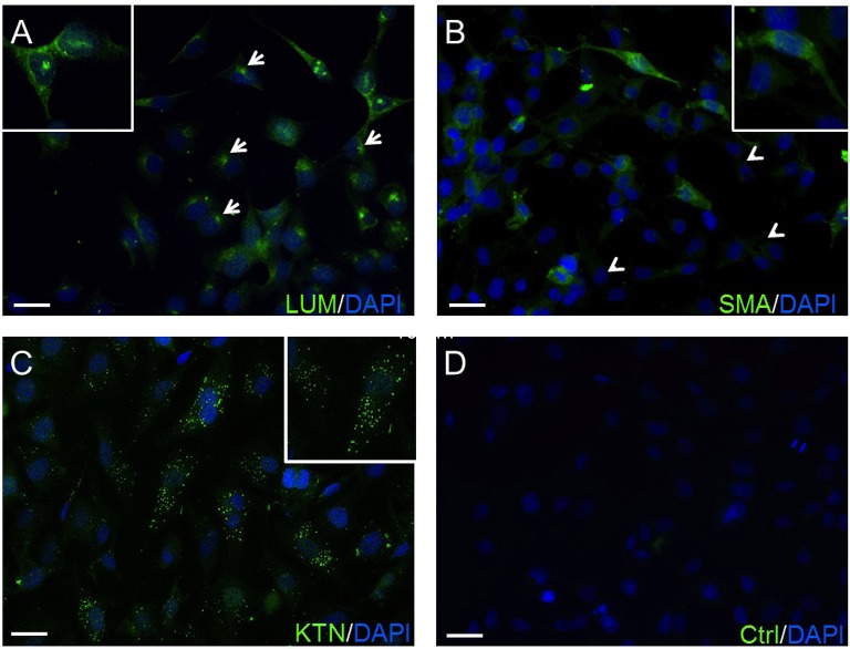
Confirmation of HCK identity. (A) The green anti-LUM antibody immunofluorescent (IF) staining is delimited in the endoplasmic reticulum (arrows). (B) Anti-α-SMA antibody IF staining pattern. Their green staining pattern is less intense. Approximately 20–30% of the HCK cells have no IF signal (arrow heads). (C) IF green staining with anti-KTN antibody detected in all HCKs. (D) A negative control (Ctrl) omitted primary antibody and does not provide a specific IF signal. In all experiments, cell nuclei were counterstained with DAPI (blue) and pictures were merged. Pictures are representative of IF results from five different assays (n = 5).

