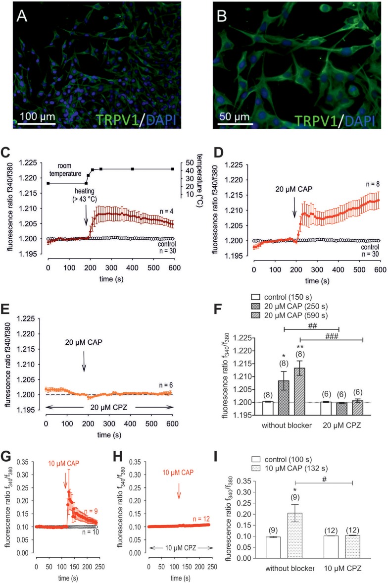
Figure 4.
Confirmation of protein and functional TRPV1 expression in cultivated human corneal keratocytes (HCK). (A,B) Localization of TRPV1 in SV40-immortalized human corneal keratocytes (HCK). IF analysis reveal subcellular TRPV1 expression (green) in all HCK cells. Cell nuclei were counterstained with DAPI (blue) and pictures were merged. Pictures are representative of IF results from different cell passages. (C) Temperature increase from ≈ 23°C to > 43°C increased [Ca2+]i (n = 4). The corresponding temperature time course is shown above the Ca2+ traces. The thermal and pharmacological changes were carried out at the time points indicated by arrows. (D) CAP (20 μM) induced an irreversible increase in Ca2+ influx (n = 8) whereas non-treated control cells maintained a constant Ca2+ baseline (n = 30). (E) Same experiment as shown in (D), but in the presence of capsazepine (CPZ). CPZ (20 μM) suppressed the CAP-induced Ca2+ increase (n = 6). (F) Summary of the experiments with CAP and heat stimulation. The asterisks (*) designate significant increases in [Ca2+]i with CAP (n = 8; p < 0.05 at the minimum; paired tested). The hashtags (#) indicate statistically significant differences in fluorescence ratios between CAP with and without CPZ (n = 6–8; p < 0.01 at the minimum; non-paired tested). (G) CAP (10 μM) induced a reversible increase in Ca2+ influx (n = 9) whereas non-treated control cells maintained a constant Ca2+ baseline (n = 10). (H) Same experiment as shown in (G), but in the presence of capsazepine (CPZ). CPZ (10 μM) suppressed the CAP-induced Ca2+ increase (n = 12). (I) Summary of the experiments with CAP and CPZ. The asterisks (*) designate significant increases in [Ca2+]i with CAP (n = 9; p < 0.05; paired tested). The hashtag (#) denotes a statistically significant difference in fluorescence ratios between CAP with and without CPZ (n = 9–12; p < 0.05; non-paired tested).

