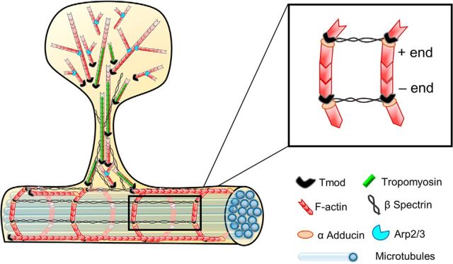
Figure 9.
Schematic representation of proposed Tmod1 and Tmod2 function in the postsynaptic compartment. Based on both our experimental data and ultrastructural and kinetic studies from other investigators, we propose that a fraction of actin filaments in the spine head is oriented with the barbed ends of the filaments toward the membrane and the pointed ends toward the spine center, or core. Tmod molecules are enriched in the spine neck and “core” of the spine head, where they cap the pointed ends of actin filaments. By contrast, Tmod molecules are rarely detected in the peripheral region of the spine head, which is characterized by newly nucleated filaments whose pointed ends are associated with the Arp2/3 complex, leading to a high number of branched filaments. βII/βIII spectrin tetramers, together with short actin filaments, form an MPS in dendrites, the spine neck, and the base of the spine head. Like βII/βIII-spectrin, Tmod immunofluorescence is detected in the spine head, but does not overlap with the PSD. The presence of Tmod in this subspine region is highly indicative of TM expression, though which TM isoform coats actin filaments in the stable core is unknown. In dendrites, short actin filaments capped by adducin at the barbed end and by Tmod2 at the pointed end, may be organized into evenly spaced rings near the plasma membrane that are connected by longitudinally oriented spectrin tetramers. Although our immunofluorescence data show that Tmod1 and Tmod2 have a largely nonoverlapping distribution in dendritic spines and along dendritic shafts, both Tmod1 and Tmod2 are presented here as “Tmod” for the sake of clarity and simplicity.

