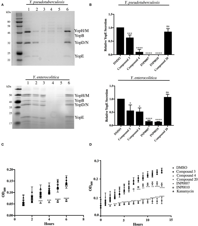
Figure 1.
Efficiency of T3SS effector protein secretion and bacterial growth in the presence of T3SS inhibitors. (A,B) The relative efficiency of effector protein secretion into the culture supernatant was analyzed following bacterial growth for 2 h under T3SS-inducing conditions in the presence of either 50 μM compound or equivalent volume of DMSO. (A) The secretome of Y. pseudotuberculosis IP2666 and Y. enterocolitica 8081 was precipitated with trichloroacetic acid, separated by SDS-PAGE, and visualized by staining with Coomassie blue. Samples were normalized to culture optical density. (1) DMSO, (2) Compound 3, (3) Compound 4, (4) INP0007, (5) INP0010, (6) Compound 20. (B) Quantification of the YopE protein band by densitometry relative to the DMSO control. The average of 3 (Y. pseudotuberculosis) or 4 (Y. enterocolitica) biological replicates ± standard deviation is shown. (C,D) Y. pseudotuberculosis IP2666 growth at 26°C in the presence of 50 μM compound or DMSO was tracked by measuring optical density. The average of three biological replicates ± standard deviation is shown and statistical significance is represented comparing compounds relative to the DMSO control. *P < 0.03; **P < 0.004; ****P < 0.0001; ***P < 0.0007 (one way ANOVA with Dunnett's post-hoc test).

