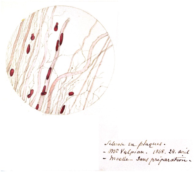
Figure 5.

Original colour drawing from Charcot’s notebook illustrating the centre of a sclerotic plaque. In the centre of the preparation is a blood vessel covered with many carmin positive nuclei and on each side are demyelinated axons of different diameter, lightly stained with carmin. In between the axons are thin fibrils, some associated with a nucleus. On the right is Charcot’s handwritten legend: Sclérose en plaques—Mr Vulpian 24 April 1868 [indicating that it was Vulpian’s patient]—Spinal cord no preparation. Source: Département de Neuropathologie, Groupe Hospitalier Pitié-Salpêtrière-Charles Foix, Musée de l’Assistance Publique-Hôpitaux de Paris.
