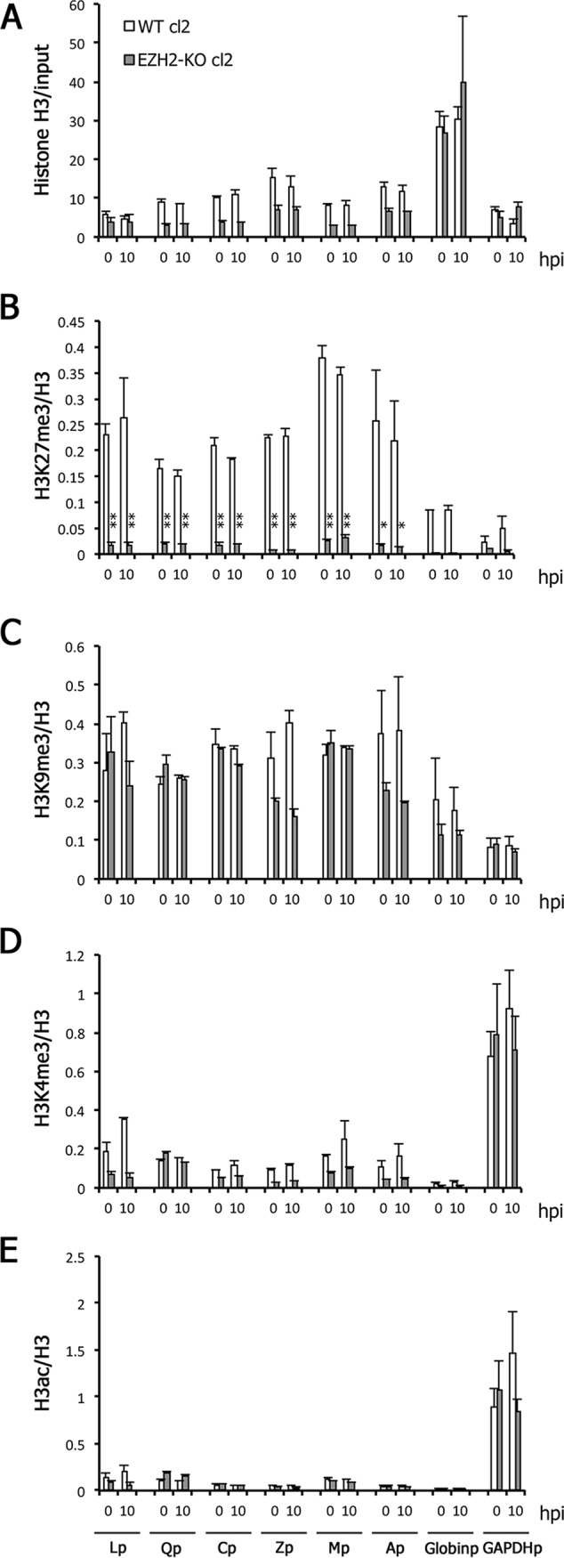
FIG 7.

Low H3K27me3 modification in the EZH2-KO cells during reactivation from latency. (A to E) WT and EZH2-KO cells latently infected with EBV (cl2) were treated with anti-IgG. Cells were harvested at 0 (latency) and 10 hpi and analyzed by ChIP using anti-histone H3 (A), anti-histone H3K27me3 (B), anti-histone H3K9me3 (C), anti-histone H3K4me3 (D), or anti-histone H3ac (E) antibodies. Data for histone H3 are shown as the percentage of the input sample (A). The data for other markers (B to E) are shown after normalization to the value of histone H3. Lp, LMP1 promoter; Qp, Q promoter; Cp, C promoter; Zp, BZLF1 promoter; Mp, BMRF1 promoter; Ap, BALF2 promoter; Globinp, Globin promoter; GAPDHp, GAPDH promoter. Student’s t test was performed. *, P < 0.05; **, P < 0.01.
