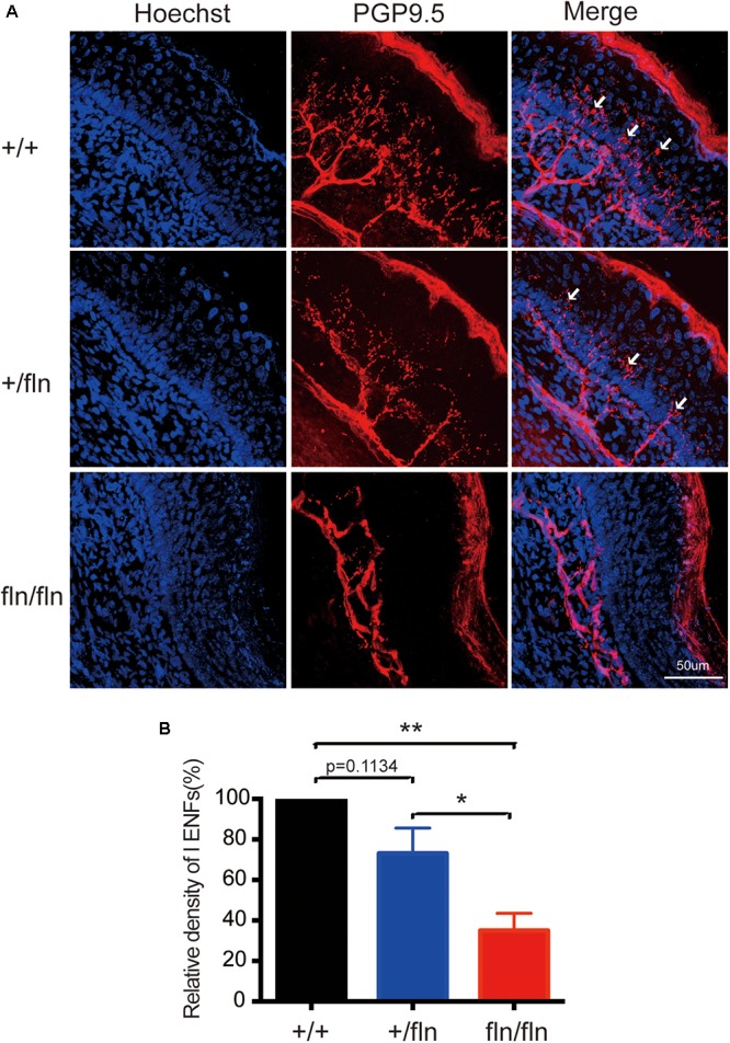
FIGURE 6.

Loss of PGP9.5-positive intra-epidermal sensory fibers (IENF) in the footpad skin in homozygous. (A) Confocal microscopic images from sections through the hindpaw footpad skin from +/+ (n = 3), +/fln (n = 3), and fln/fln (n = 3) P1 mice stained with pan-nerve fiber marker PGP9.5 (red) and nuclear marker Hoechst (blue) to visualize skin cells. The arrowhead indicates the free nerve endings, (B) Quantification of PGP9.5-positive IENF intensities. Mean ± SEM, ∗p < 0.05, ∗∗p < 0.01, one way ANOVA test followed by Tukey’s multiple comparisons test.
