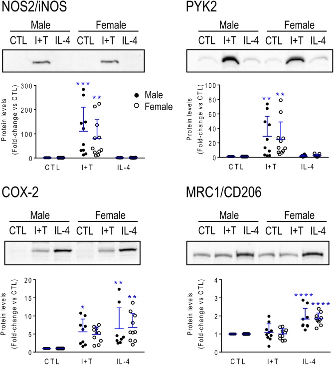
FIGURE 2.
Neonatal (P1) microglia: Exemplary pro- and anti-inflammatory proteins. Rat microglia were harvested 24 h after treatment with IFNγ + TNFα (I+T) or IL-4. (Upper) Representative Western blots for the pro-inflammatory markers, iNOS, COX-2 and PYK2, and the anti-inflammatory marker, MRC1/CD206. (Lower) Individual values show fold-changes with respect to unstimulated (control) microglia and the mean ± SD is indicated. Differences from control microglia are shown as: ∗p < 0.05; ∗∗p < 0.01; ∗∗∗p < 0.001, and ∗∗∗∗p < 0.0001.

