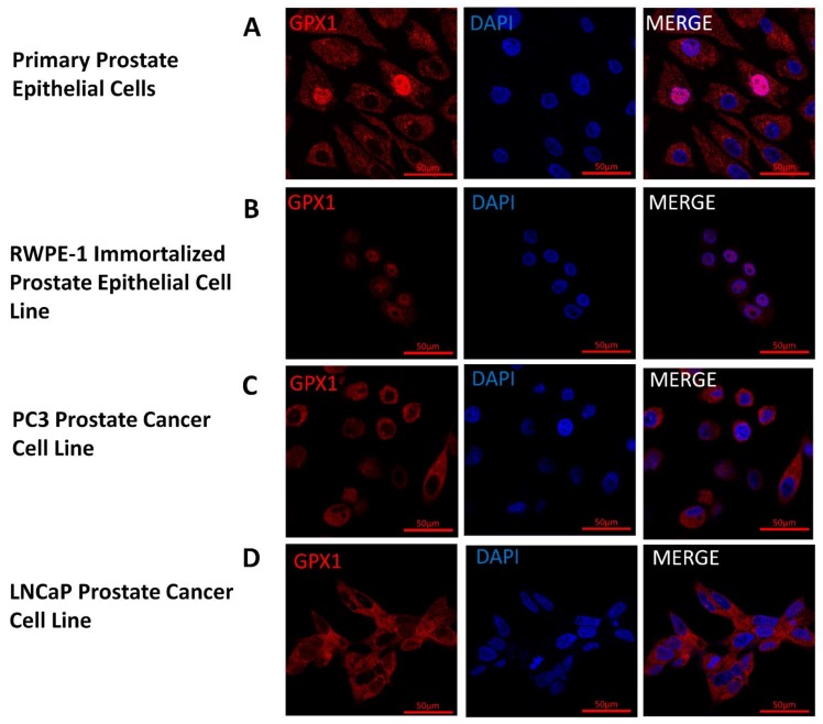
Figure 3.
Location and levels of GPX1 in cultured cells. GPX1 appears in red and nuclei are indicated by staining with 4’,6-diamidino-2-phenylindole (DAPI) in blue. (A) Primary Prostate Epithelial Cell; (B) RWPE-1 Immortalized Prostate Epithelial Cell Line; (C) PC3 Prostate Cancer Cell Line; (D) LNCaP Prostate Cancer Cell Line.

