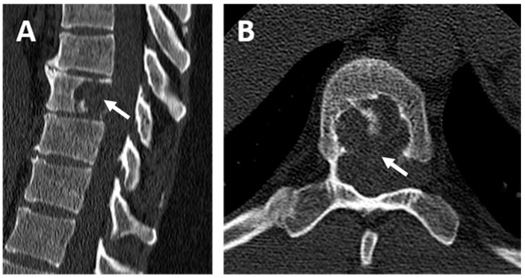
Figure 1.
Computed tomography scan (CT scan) of the patient’s spine. (A) Dorsal CT scan in the sagittal plane and (B) dorsal CT scan in the axial plane centered on the ninth dorsal vertebra (T9). Osteolytic lesion containing septa centered on the body of the ninth dorsal vertebra (white arrow) with lysis of the posterior wall without osteo-condensation and fluid density.

