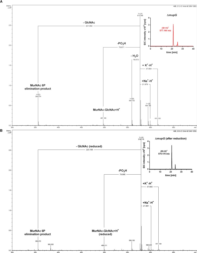
FIGURE 3.
Accumulation of MurNAc 6P-GlcNAc in ΔmupG cells. S. aureus ΔmupG was grown in LB medium to stationary phase (24 h). Acetone extracts of the cytosolic fractions were analyzed by LC-MS in positive ion-mode, before (A) and after (B) NaBH4 reduction. (A) Shown are the fragmentation patterns obtained by in-source decay of the compound that accumulates specifically in ΔmupG mutant cells and eluates at a retention time of 21.2–21.4 min, represented as extracted ion chromatogram with an observed mass of (M+H)+ = 577.166 m/z (EIC × 105 counts per s [cps], upper right corner). Representative mass spectra from three biological replicates are illustrated. (B) Shown are the fragmentation patterns after reduction of the sample with NaBH4, which results in the formation of a new compound that elutes at 21.0 min, represented by an EIC with an observed mass of (M+H)+ = 579.178 m/z (upper right corner). The mass spectra fragmentation patterns of both compounds were presented in Compass DataAnalysis program (Bruker) from 300 to 700 m/z. The obtained fragmentation pattern indicated that the substrate for MupG is MurNAc 6P-GlcNAc.

