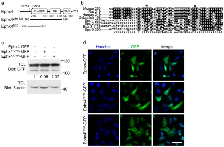
Figure 1.
Generation of putative mutants of Ephexin4 that disrupt its intermolecular interaction. (a) Schematic diagram of the constructs used in this study. Ephx4, Ephexin4. (b) Amino acid sequence alignment of Ephexin proteins. Sequences were aligned using ClustalW and displayed using BoxShade. Asterisks indicate highly conserved residues in Ephexin proteins that were mutated in this study. (c) 293T cells were transfected with the indicated plasmids. Two days later, cells were lysed and proteins in the lysates were detected by immunoblotting. TCL, total cell lysate. n = 4. (d) LR73 cells were transfected with the indicated plasmids. GFP signals indicating expression of the transfected plasmids were observed by microscopy. Scale bar, 40 µm. n = 3. Images shown are representative of at least three independent experiments.

