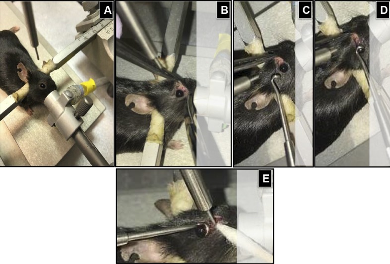
Figure 2.

The procedure for controlled ocular impact. (A) The anesthesia was maintained through an inlet tube mounted to a cylindric metallic shield covering the bite plate; (B) by using a pair of blunt laminectomy forceps and scissors, an incision was made at the medial canthus; (C–E) using a noninvasive approach, the eyeball was retracted from the orbital margin; leaving extraocular tissues fully exposed to the controlled impaction from a blunt impactor tip 1 mm in diameter.
