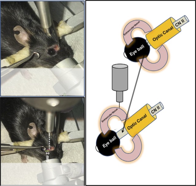
Figure 3.
Schematic presentation of the injury site at a distance of 2 to 3 mm from the posterior pole of the globe. The retraction of the eyeball from the orbital margin makes the intraorbital portion of the optic nerve easily accessible and reproducibly injured by the application of controlled impact to the extraocular tissue in the orbital area posterior to the globe.

