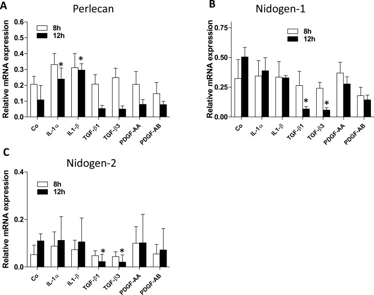
Figure 1.
EBM component mRNA expression in primary cultures of rabbit keratocytes in presence of different cytokines/growth factors. Keratocan+ keratocytes were cultured and treated with 10 ng/mL IL-1α, 10 ng/mL IL-1β, 2 ng/mL TGF-β1, 10 ng/mL TGF-β3, 10 ng/mL PDGF-AA, or 10 ng/mL PDGF-AB for 8 or 12 hours. Expression of perlecan (A), nidogen-1 (B), and nidogen-2 (C) mRNA was measured by qRT-PCR and normalized to 18S rRNA as described in the material and methods section. “Co” represents primary cultured keratocan + keratocytes in the medium without added cytokines or growth factors. Data for each BM component and each cytokine or growth factor are presented as means of three independent experiments and statistical comparisons were made between vehicle-treated control keratocytes and cytokine- or growth factor–treated keratocytes at the same time points. No comparisons were made between the 8- and 12-hour time points.

