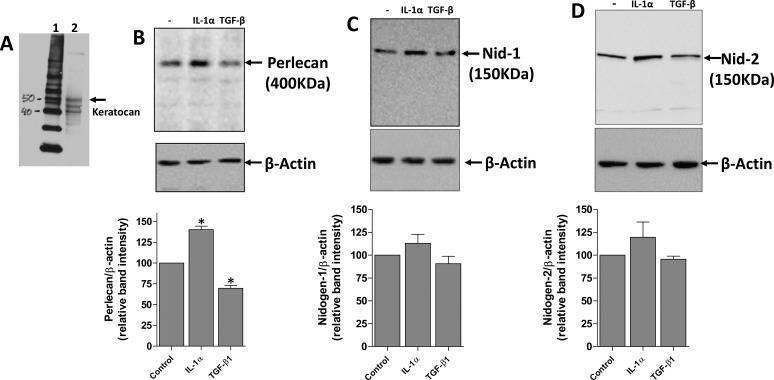
Figure 2.
Regulation of EBM component protein expression by IL-1α and TGF-β1 in primary rabbit keratocytes. Primary keratocan+ keratocytes were cultured and treated with 10 ng/mL IL-1α, 2 ng/mL TGF-β1, or left untreated for 16 hours. Keratocytes to be used in the experiments were lysed and keratocan of the expected size (50 kDa45) was detected (A) to confirm these cells were keratocan+ keratocytes at the beginning of the exposure. (B) Perlecan, (C) nidogen-1, and (D) nidogen-2 expression detected by Western blot. Cell extracts used for perlecan Western blots were treated with heparitinase III, as was described in the methods. β-actin was used as a loading control for each experiment. A representative Western blot of the three performed for each BM component is shown. The graphs beneath each Western blot was obtained by densitometry analysis of the bands from each of the three Western blots from different experiments. *The change in BM protein was statistically significant (P < 0.05) compared with the control keratocytes.

