Figure 5.
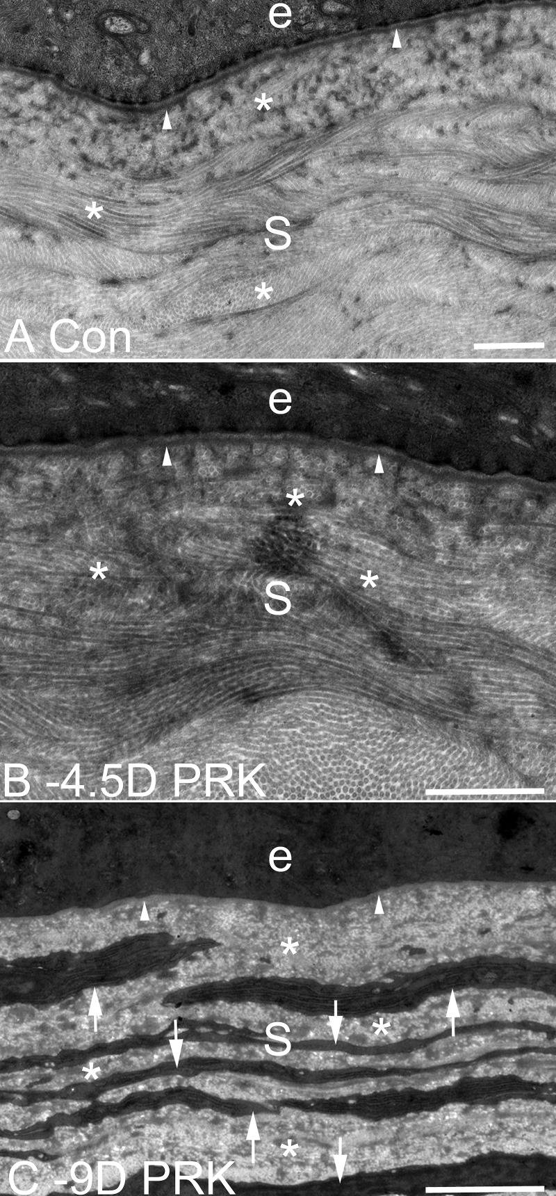
TEM of EBM ultrastructure and the anterior stroma at 1 month after −9- or −4.5-D PRK, or in control unwounded, rabbit corneas. (A) In an unwounded cornea, lamina lucida, and lamina densa (arrowheads) are present beneath the epithelium (e). S is stroma in all panels. (B) At 1 month after −4.5-D PRK, the normal lamina lucida and lamina densa (arrowheads) of the EBM are regenerated beneath the epithelium (e). There are no cells seen in the subepithelial stroma with high levels of rough endoplasmic reticulum suggestive of myofibroblasts. *In panels (A) and (B), note the uniform diameter of collagen fibrils throughout the stroma, with some seen longitudinally and others cut transversely. (C) At 1 month after −9-D PRK, normal lamina lucida and lamina densa cannot be detected (arrowheads) beneath the epithelium (e) and the subepithelial stromal is filled with layered myofibroblasts (arrows, and same cells seen in IHC for α-SMA shown in [C]) with large amounts of rough endoplasmic reticulum. Note the relative disorganization of the collagen fibrils in the extracellular matrix (*) between the myofibroblast cells compared with the fibrils with uniform thickness and regular distribution in the stroma of panels (A) and (B). Magnification bars are 1 μm in each panel.

