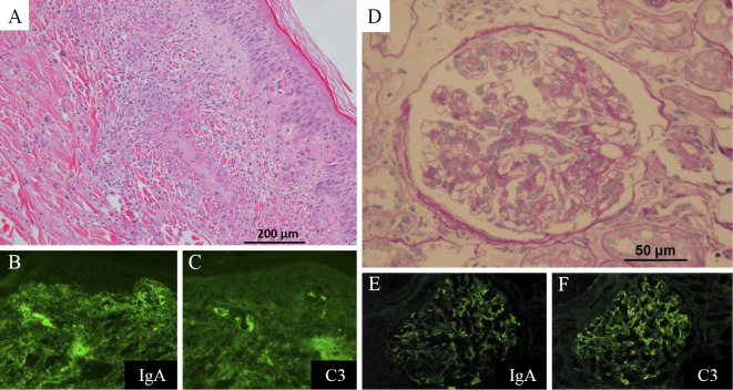
Figure 3.
(A) Histopathological findings of the skin specimen showed leukocytoclastic vasculitis in the small vessels throughout the dermis (Hematoxylin and Eosin staining). (B), (C) An immunofluorescence study showing perivascular deposits of immunoglobulin A (IgA) and C3. (D) A renal biopsy specimen showed endocapillary proliferative glomerulonephritis (PAS stain). (E), (F) An immunofluorescence study indicated mesangial IgA and C3 deposition.

