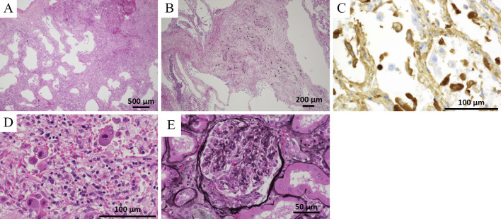
Figure 6.
(A) The histopathological findings of the postmortem specimen showed erythrocyte extravasation and leukocyte infiltration in the interstitium of the lung (Hematoxylin and Eosin (H&E) staining). Leukocytoclastic vasculitis was not seen. (B) Fibrous thickening and pneumoconiosis nodules were seen around the bronchioles of the bilateral upper lungs (H&E staining). (C) An immunohistochemical examination showed that IgA was positive in the alveolar walls. (D) Cytomegalic inclusion bodies (owl’s eye) were also seen in the lung specimen (H&E staining). (E) There were crescent bodies in several glomeruli that had not been seen antemortem (PAM stain).

