Fig. 7.
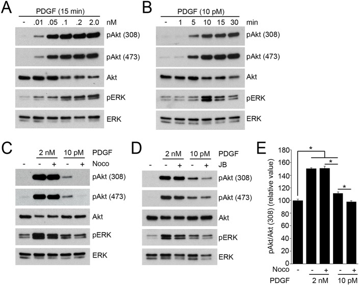
Low concentrations of PDGF induce cytoskeleton-dependent Akt phosphorylation. (A) Dose-dependent assay of PDGF stimulation. At 15 min following stimulation with PDGF (0.01–2.0 nM), MEFs were lysed for biochemical analysis. The intensity of pAkt induced by PDGF decreased at 0.01 nM (10 pM). (B) Time course of signaling in response to 10 pM PDGF. Akt phosphorylation was prolonged but ERK phosphorylation was transient. (C) Nocodazole (Noco) treatment attenuated pAkt(308) and pAkt(473) induced by 10 pM PDGF, but not by 2 nM PDGF. (D) J/B treatment attenuated pAkt(308) and pAkt(473) induced by 10 pM PDGF, but not by 2 nM PDGF. (E) Quantification of immunofluorescence images of pAkt(308) and Akt. MEFs were stimulated by PDGF (2 nM or 10 pM) for 3 min with or without nocodazole. Ratio images of pAkt(308) and Akt were calculated to quantify the intensity of pAkt in each condition. The relative ratio value of 10 pM PDGF-stimulated cells was significantly higher than for untreated cells and was attenuated by nocodazole. The values for cells stimulated with 2 nM PDGF were not attenuated by nocodazole. More than 15 cells were observed for each condition. One-way ANOVA was applied for the statistics; *P<0.05.

