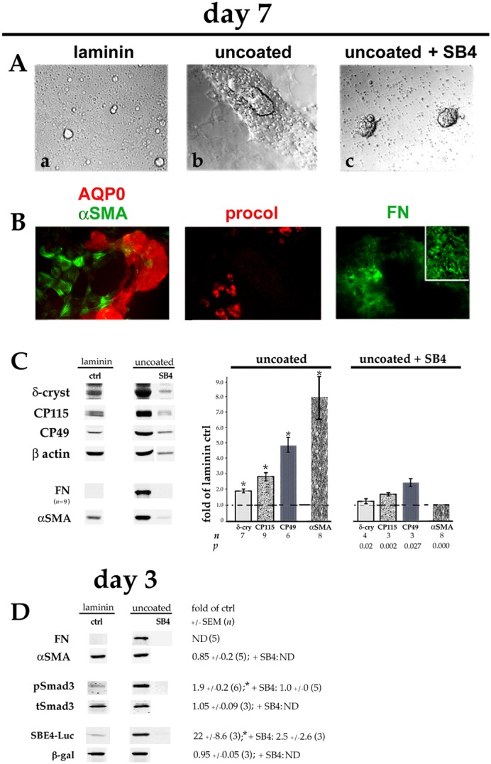
Fig. 5.

Plating DCDMLs on uncoated tissue culture plastic upregulates EMyT, fiber cell differentiation and TGFβ signaling. DCDMLs were plated in uncoated tissue culture wells (or, as a control, in laminin-coated wells) at 1.2×10⁵ cells/96 plate well on day 0. Cells were cultured from day 1 in M199/BOTS with or without SB431542 (SB4) as indicated. (A) Phase-contrast images of cultures on day 7; (a,b) show near-confluent regions, whereas (c) shows two balls of tightly packed cells surrounded by phase-bright cell blebs. (B) Cultures plated on uncoated plastic were immunostained for aquaporin-0 (AQP0) and the fibrotic markers αSMA, procollagen 1 (procol), and FN. Near-confluent regions of cultures are shown; inset is a high-magnification view of NaOH-insoluble FN extracellular fibrils. All markers assessed in a minimum of three independent experiments with similar results. (C) Cells were assayed on day 7 for expression of fiber cell and EMT/EMyT markers as in Fig. 4. Note that the lower number of adherent cells in cultures plated on uncoated TC plastic and treated with SB431542 resulted in lower levels of β-actin per sample. (D) Cultures were analyzed on day 3 for FN and αSMA, or for pSmad3, total Smad3 (tSmad3) and SBE4–luciferase (SBE4–Luc) expression using quantitative western blot as in Fig. 1. *P≤0.001 compared to untreated cells plated on laminin (ctrl); all other data sets not significantly different from control (P>0.2) unless indicated otherwise. ND, not determined.
