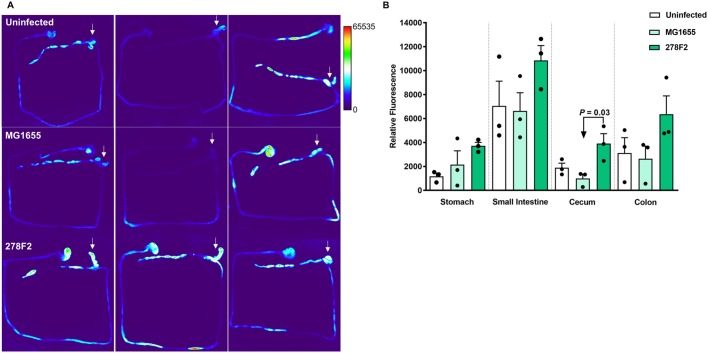
Fig. 3.
Evaluation of mucosal inflammation in mice with a microbiota consisting of the altered Schaedler flora (ASF). Mice received a tail vein injection with 2 nmol per mouse of ProSense 680 and were imaged 18 h later using the In Vivo Multispectral Imaging System FX Pro. (A) The entire GI tract was excised from uninfected (top row), non-pathogenic E. coli MG1655-inoculated (middle row), and enterohemorrhagic E. coli (EHEC) 278F2-inoculated (bottom row) mice (n=3 per group). Arrows indicate the cecum. (B) Relative fluorescence was measured in ImageJ. An ANOVA followed by Tukey's method for multiple means comparison was used to compare total corrected cellular fluorescence of intestinal sections. Each symbol represents an individual animal and bars represent mean±s.e.m.

