Fig. 4.
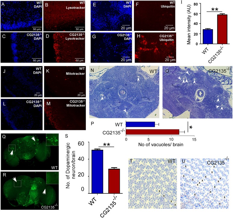
Neuropathological abnormalities in the CG2135−/− fly. (A-D) LysoTracker (red) and DAPI (blue) staining of 30-day-old fly brains, showing increased abundance of the LysoTracker-positive vesicles in the CG2135−/− fly compared to the wild type (WT). (E-H) Immunostaining of the 30-day-old fly brains with anti-ubiquitin antibody (red), showing an elevated level of ubiquitinated proteins in the CG2135−/− fly. (I) Bar graph showing a significant increase in mean intensity of ubiquitin staining in the CG2135−/− fly brain. (J-M) MitoTracker (red) and DAPI (blue) staining of the 30-day-old fly brains, showing increased mitochondrial accumulation in the CG2135−/− fly brain. (N,O) Toluidine-Blue-stained brain sections of the 30-day-old flies, showing increased vacuolation (indicated by arrowheads) in the CG2135−/− fly. (P) The number of vacuoles in the WT and CG2135−/− fly brain sections. (Q,R) Immunostaining of the 30-day-old fly brains with anti-tyrosine-hydroxylase antibody (green), showing loss of dopaminergic neurons in the CG2135−/− fly brain compared to the WT flies. Enlarged view of a dopaminergic neuron cell body from the boxed regions of the brain is provided in the inset. (S) Quantitation of dopaminergic neurons in the WT and CG2135−/− fly brains. (T,U) Toluidine-Blue-stained retinal sections showing well-preserved ommatidial architectures in the 30-day-old WT fly, as opposed to defective ommatidial organization (‘x’ showing the gaps) in the age-matched CG2135−/− fly. Error bar represents s.e.m. of values from at least three independent experiments. *P≤0.05, **P≤0.01.

