Fig. 5.
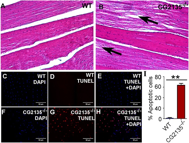
Muscle degeneration in the CG2135−/− fly. (A,B) Hematoxylin and eosin staining of longitudinal sections of thoracic muscle of 30-day-old flies, showing intact muscle structure in the wild type (WT) fly as opposed to fragmented muscle fibres (indicated by arrows) in the CG2135−/− fly. (C-H) TUNEL staining (red) of the thoracic muscles, showing widespread apoptosis in the 30-day-old CG2135−/− fly. The total number of cells is represented by DAPI (blue). (I) Bar graph showing that the percentage of TUNEL-positive (apoptotic) cells in the muscle of the CG2135−/− fly is significantly higher than in the WT fly. Error bars represent the s.e.m. of values from four independent experiments. **P≤0.01.

