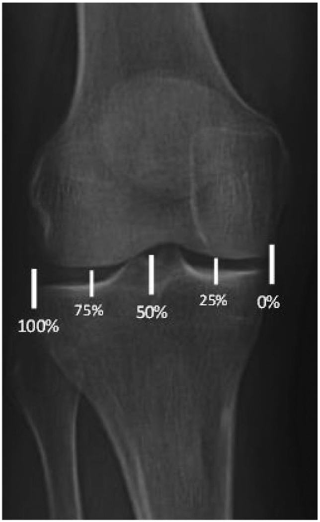
Figure 1.

Weight-bearing alignment as described by Noyes et al. [10–12,25]. The medial edge is designated as 0% and the lateral edge as 100%. A line is drawn from the center of the femoral head to the center of the tibiotalar joint and is measured at the point where the line intersects the tibial plateau.
