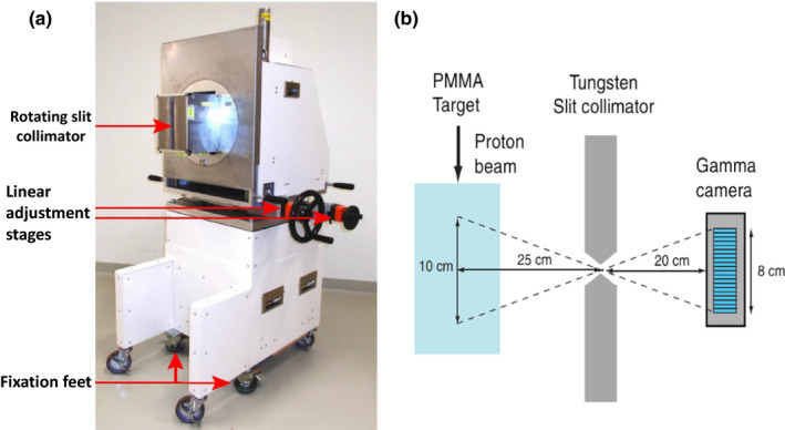
Figure 5.

(a) The clinical slit camera used for patient range verification (adapted from Richter et al.85) as well as (b) a schematic drawing of the slit‐collimator imaging concept in which the originating position of the PG is derived from the vector connecting its point of detection in the camera and the opening of the collimator (adapted from Perali et al.80), with permission.
