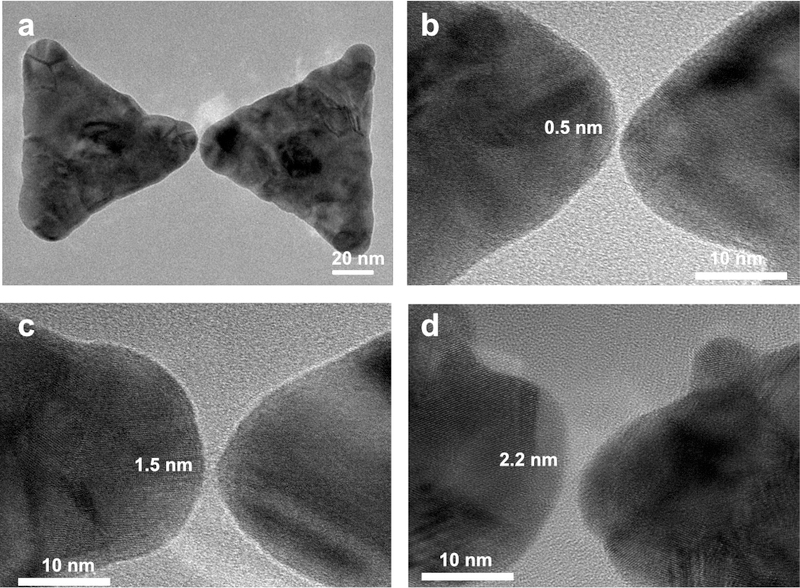
Figure 3. TEM images of bowtie structures with different gap-sizes.

(a) TEM image of a typical bowtie structure with an ultra-small nanogap. (b-d) Close-up TEM images of bowtie structure with 0.5-nm (b), 1.5-nm (c), and 2.2-nm (d) nanogaps.

(a) TEM image of a typical bowtie structure with an ultra-small nanogap. (b-d) Close-up TEM images of bowtie structure with 0.5-nm (b), 1.5-nm (c), and 2.2-nm (d) nanogaps.