Fig. 1.
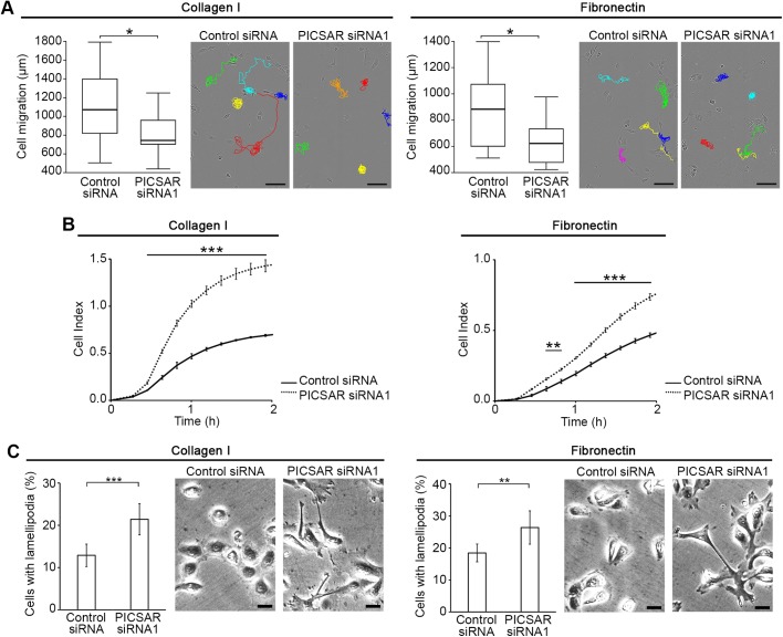
Knockdown of PICSAR inhibits cSCC cell migration and increases cSCC cell adhesion and formation of lamellipodia. cSCC cells (UT-SCC12A) were transfected with PICSAR siRNA (siRNA1) or control siRNA. (A) Cells were plated on collagen I or fibronectin 72 h after transfection and migration of individual cells was imaged using the IncuCyte ZOOM® real-time cell imaging system. Cell tracking (n=15) was quantitated using ImageJ software. Median±s.d. is shown; *P<0.05, Mann–Whitney two-way U-test. Representative images of cell tracking are shown. Scale bars: 100 µm. (B) Cells were plated on collagen I or fibronectin coated 96-well electronic microtiter plate 72 h after transfection and cell adhesion was measured using the xCELLigence system (n=3). The cell index indicates the readout of the microelectrode impedance which corresponds to the strength of cell adhesion. Mean±s.d. is shown; **P<0.01, ***P<0.001; two-tailed t-test. (C) Cells were plated on collagen I or fibronectin 72 h after transfection and fixed 4 h after plating. The number of lamellipodia containing cells was quantitated from microscopic images with a 20x objective (n=3). Representative images of the quantification are shown. Scale bars: 10 µm. Mean±s.d. is shown; **P<0.01, ***P<0.001; two-tailed t-test.

