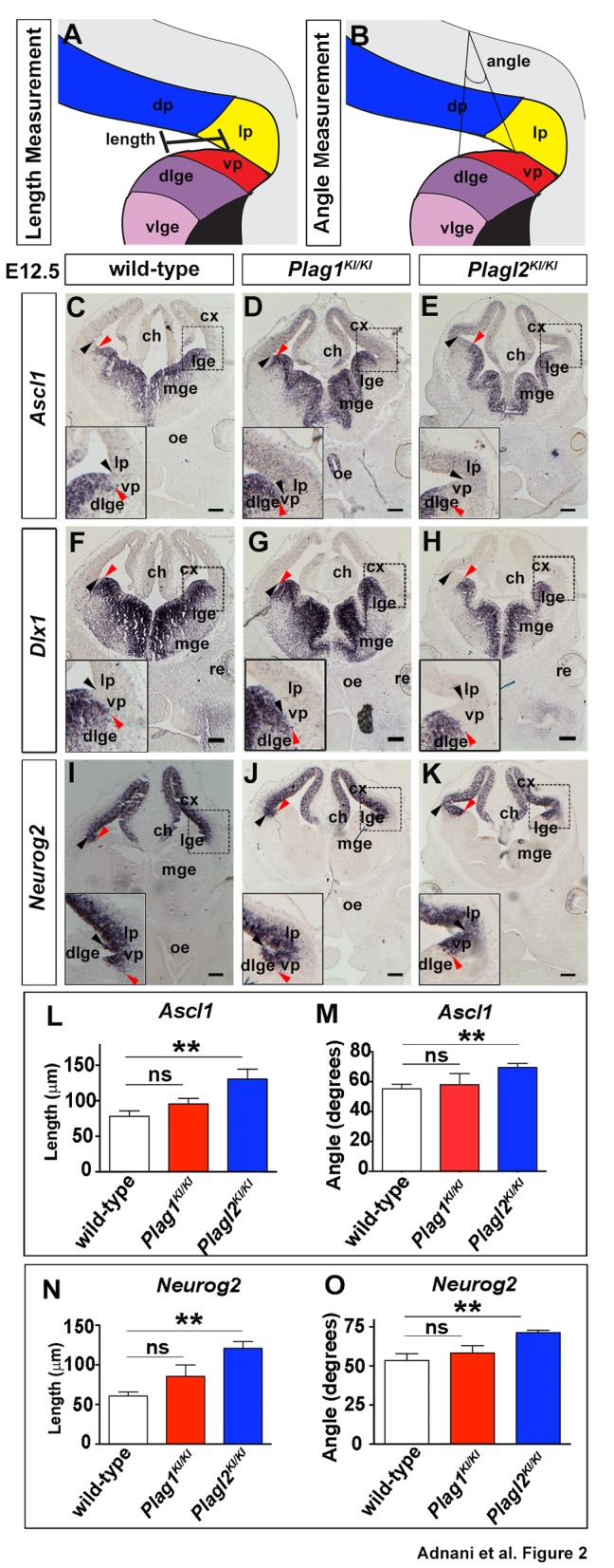
Fig. 2.

Plag1 and Plagl2 are required to pattern the embryonic telencephalon. (A,B) Schematic representations of length (A) and angle (B) measurements of the ventral pallium, extending from the corticostriatal angle to the gene expression border. (C–K) Expression of Ascl1 (C–E), Dlx1 (F–H) and Neurog2 (I–K) in E12.5 wild-type (C,F,I), Plag1KI/KI (D,G,J) and Plagl2KI/KI (E,H,K) brains. Black arrowheads mark the corticostriatal angle and red arrowheads mark the ventral pallial gene expression limit. (L–O) Quantification of the length (L,N) and angle (M,O) of the ventral pallium based on the expression of Ascl1 (L,M), and Neurog2 (N,O). Error bars are s.e.m. ns, not significant; *P<0.05, **P<0.01, and ***P<0.005. ch, cortical hem; cx, neocortex; dlge, dorsal lateral ganglionic eminence; dp, dorsal pallium; lge, lateral ganglionic eminence; lp, lateral pallium; mge, medial ganglionic eminence; mp, medial pallium; oe, olfactory epithelium; re, retina; vlge, ventral lateral ganglionic eminence; vp, ventral pallium. Scale bars: 250 μm.
