Fig. 4.
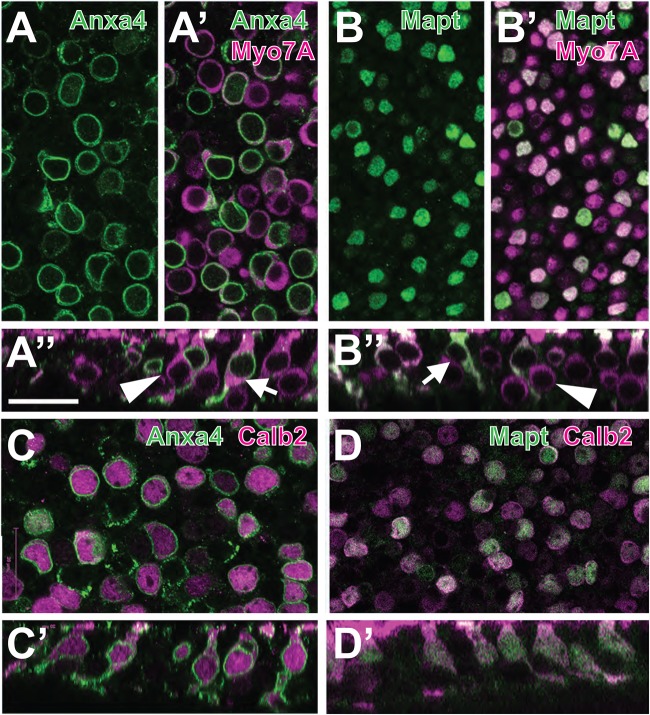
Anxa4 and Mapt are markers for Type II HCs. (A) Surface view from a P64 utricle labeled with anti-Anxa4 (green). Anxa4 is expressed along the cell and nuclear membranes of a subset of cells. (A′) The same view as in A, but also showing expression of the pan-HC marker Myo7a in magenta. Anxa4 co-localizes with a subset of HCs. (A″) Orthogonal view from the same sample as in A. Arrow indicates an Anxa4-positive cell. Note the more lumenal position of the nucleus and the wider HC neck. By comparison, an Anxa4-negative cell (arrowhead) has a more basal nucleus and a narrow neck. (B–B″). Similar views as those in A, except with expression of Mapt illustrated in green. Arrow and Arrowhead in B″ illustrate similar differences in the morphologies of Mapt+ and Mapt− cells. (C,D) To confirm that Anxa4 and Mapt are markers of Type II HCs, P62 utricles were double-labeled with Anxa4 (C) or Mapt (D) and the Type II HC marker, Calb2. Note co-labeling of both Anxa4 and Mapt with Calb2. Scale bar in A″, same for all other panels: 20 µm.

