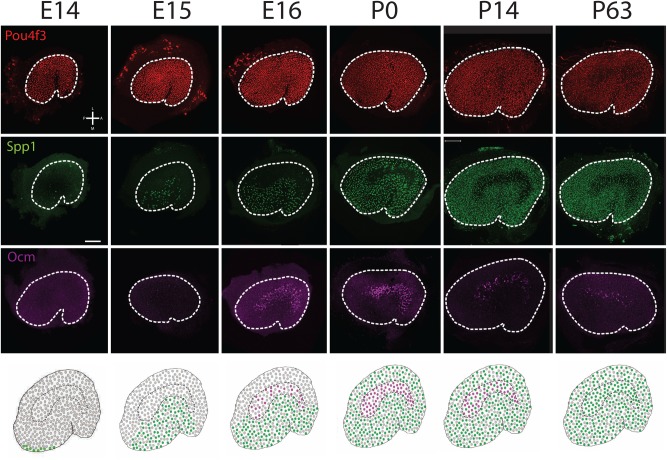
Fig. 8.
Temporal and spatial development of Type I HCs. Low magnification surface images of entire utricles obtained at the indicated time points. The upper three rows show a pan specific HC label (Pou4f3 in red), and the Type I HC markers Spp1 (green) or Ocm (magenta). The bottom row illustrates a cartoon summary of Spp1 (green) and Ocm (magenta) expression at each age. At E14, while the utricle contains a uniform distribution of HCs, Spp1+ HCs, which are predominantly Type Is, are restricted to the extreme medial-posterior region of the epithelium. At E15, Spp1+ cells have extended anteriorly and laterally. At E16, Spp1+ cells are present in the entire medial half of the utricle and Ocm+ HCs are present in the striola. At P0, cells that express Spp1 are present throughout the utricle with the exception of the striolar HCs which still express Ocm. At P14, Spp1+ Type I HCs are now present in the striola while expression of Ocm has decreased. At P63, Ocm+ HCs are almost completely absent, while most striolar Type I HCs are now positive for Spp1. Orientation for all images is indicated in the upper left hand panel. Scale bar: 100 µm.

