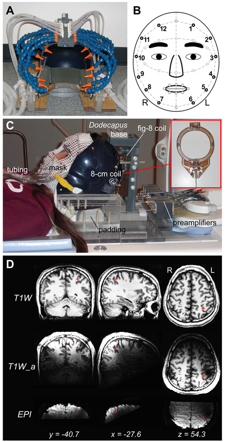
Figure 1.
MRI-compatible devices for radio-frequency signal acquisition and tactile stimulation. (A) Dodecapus manifold mounted above a baseball helmet for Experiment 1. (B) A schematic of stimulation sites (black circles) on the face in both experiments. (C) Integration of wearable tactile stimulation (mask) and surface coils for Experiment 2. (D) Location of the parietal face area (red cross) in the left hemisphere of Subject 1. T1W: T1-weighted structural images (average of two image sets); T1W_a: T1-weighted alignment images; EPI: functional images.

