
Figure 6.
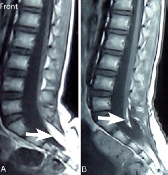
Typical case.
(A) T1-weighted magnetic resonance image of a 7-year-old female patient with transitional lipoma at S1–3 level. White arrow shows lipoma. (B) T1-weighted magnetic resonance image scanned 6 months after surgery. White arrow shows that the lipoma was almost completely removed.