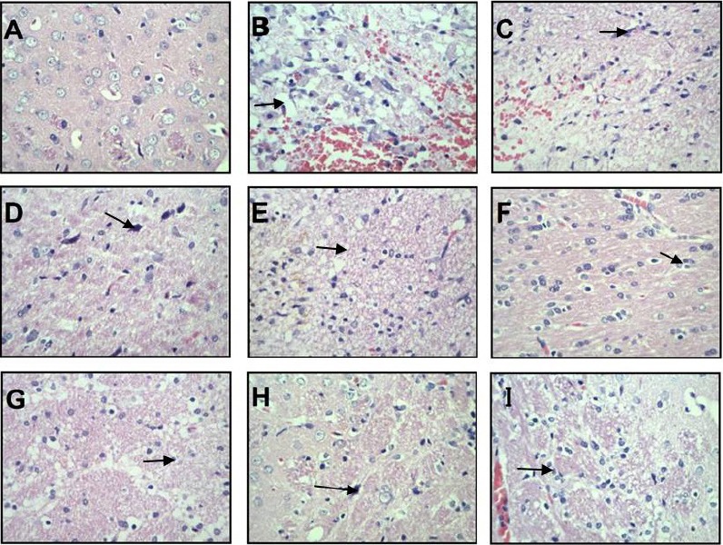
Figure 1.

Hematoxylin-eosin staining of brain tissue from each group (light microscopy).
(A) Sham-operation group; (B–E): 3-, 9-, 24-, and 48-hour model groups; (F–I): 3-, 9-, 24-, and 48-hour acupuncture groups. Arrows point to cysts (B), inflammatory cell infiltration (C), nuclear pyknosis (D), irregular cysts (E), nuclei without distinct changes (F), smaller neurons (G), edema in relatively small regions (H), and severe edema (I). Original magnification, 400×.
