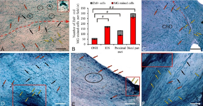
Figure 1.

Localization of neurolin (a regeneration-associated, Zn-8 labeled protein) in the contralateral optic nerve of rainbow trout, Oncorhynchus mykiss, at 1 week after unilateral eye injury (UEI).
(A) Immunolabeling of Zn8 in the middle part of the contralateral nerve: Zn8+ cells (indicated by red arrows), Zn8– rounded cells (white arrows), Zn8– elongated cells (black arrows), Zn8+ axons (yellow arrows), and Zn8+ cells and clasters (inset) in the central part of the optic nerve (outlined by ovals); the red dotted line delimits the middle portion of the nerve and shows the direction of cell migration. (B) Ratio of Zn8+ and methyl green (MG) stained cells (mean ± SD) in the optic nerve head (ONH), intraorbital segment (IOS), proximal and distal parts of the contralateral nerve (n = 5 in each group; #P < 0.05, ##P < 0.01; one-way analysis of variance (ANOVA) followed by the Student-Newman-Keuls post hoc test); the error bars correspond to the standard deviation. (C) Immunolabeling of Zn8 in the optic chiasma (arrow indications here and below see in A). (D) Immunolabeling of Zn8 in the IOS with an aggregation of germinating Zn8+ axons (in red rectangle). (E) Immunolabeling of Zn8 in the proximal part of the contralateral nerve with an aggregation of germinating axons (in the red rectangle) and aggregation of Zn8– astrocytes (in black oval). (F) Immunolabeling of Zn8 in the distal part of the contralateral nerve; degenerating Zn8– fibers are indicated by black arrows (other arrow indications see above). Peroxidase Zn8– immunolabeling on optic nerve sections with methyl green staining. Scale bars: A, 100 μm; C–F, 50 μm.
